By Lynn Anderson Davy
UI Strategic Communication
Training orthopedic surgeons to repair fractured bones can take years and cost hundreds of thousands of dollars. A University of Iowa research team wants to improve the process with a sophisticated, but portable, orthopedic surgery simulator that uses small cameras and synthetic bones to mimic surgery and provide residents with immediate feedback.
In most cases, doctors training in orthopedics as part of a residency program work with more experienced physicians in the operating room (OR). Although helpful, the process offers little opportunity for less experienced doctors to do much more than observe or assist. And when residents first pick up a scalpel to perform a surgery themselves, it can be distracting to have another doctor coaching or advising from the sidelines.
“Imagine you’re in the OR with a patient on the operating table and someone is beside you saying, ‘Drop your hand a bit’ or ‘That’s right, but go more slowly here,” says Geb Thomas, a professor of mechanical and industrial engineering in the UI College of Engineering. “So, our simulator came out of this idea of improving training and building skills even before a resident gets into the OR.”
For several years, Thomas and research partner Don Anderson, a biomedical engineer and professor of orthopedics and rehabilitation in the UI Roy J. and Lucille A. Carver College of Medicine, have been developing the simulator with a team of specialists and student researchers—even spinning off a small company to market the device. Now they’re focused on testing whether it improves performance in the OR.
“At this stage, we need to be able to scientifically assess if a resident’s ability to perform a surgical procedure has improved as a result of his or her time working with the simulator,” Anderson says. “This means studying fluoroscopic images to quantitatively determine the accuracy of the wire placement, and then comparing residents who participated in our training with those who haven’t. With this information, we’ll have a good idea if our simulator is helping or not.”
Potential uses for the simulator are many, and the project has won the support of the American Board of Orthopaedic Surgery and the Orthopaedic Trauma Association. These groups are eager to see if the simulator can help improve the quality and efficiency of training while ensuring residents get practice in all sorts of surgeries.
In some cases, hospitals use virtual reality training programs to help residents gain surgical expertise, but these experiences can’t replicate the feel of drilling through bone. “What we are interested in doing is getting real tools in a surgeon’s hands rather than a game,” Anderson says.
The federal government also has expressed interest in the device. Recently, the Agency for Healthcare Research and Quality, a part of the U.S. Department of Health and Human Services that invests in research to make health care safer and improve quality, awarded Thomas and Anderson a five-year, $2 million grant to assess surgical performance improvement at Midwest university hospitals, as well as to implement training on the simulator at national fracture courses.
“It’s pretty exciting because we have colleagues from hospitals in other parts of the country who are begging us to include them in the research so their residents can also benefit from training on the simulator,” Anderson says. “So far, we’ve trained residents at the University of Minnesota, the Mayo Clinic, and the University of Nebraska Medical Center.”
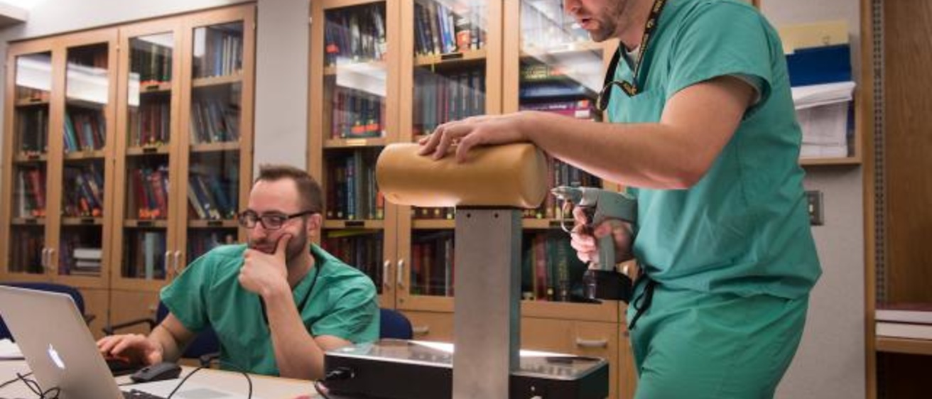
Potential uses for the simulator are many, and the project has won the support of the American Board of Orthopaedic Surgery and the Orthopaedic Trauma Association. Photo by Tim Schoon.
During recent tests at UI Hospitals and Clinics (UIHC), the hands-on experience offered by the simulator was a hit with residents. Orthopedic residents worked with UI biomedical engineering graduate student Steven Long who, along with several other graduate and undergraduate students, worked with the research team to perfect the simulator. Long explained how to use the simulator and inserted new foam bones into the simulator when necessary. On this day, residents used the orthopedic simulator to practice repairing a fractured femur.
Although this type of surgery is routine, it is far from simple. It requires drilling a long wire into the femur and guiding it to the correct spot at the apex of the femoral head, where the femur articulates in the pelvis. During surgery, doctors use fluoroscopy snapshots to make sure they are inserting the wire correctly. The trick, however, is to use as few snapshots as possible to save time and reduce radiation exposure.
During one session on the simulator, Alan Shamrock, a resident in UIHC’s Department of Orthopedics and Rehabilitation, worked slowly, changing drill angles as he attempted to correctly place the wire in the femur. Working with him was fellow orthopedics resident Chris Lindsay, who operated a simulated fluoroscopy machine to provide Shamrock with the images (simulated and presented on a laptop screen) he needed to guide the wire through the fake femur.
As Shamrock drilled, he periodically stopped and asked Lindsay to take a fluoroscopic image. “Shoot that,” he said, each time he needed to confirm the position of the wire inside the bone. It took several tries and several dozen pretend snapshots before Shamrock was able to get the wire in the correct position. When he was finished, he reviewed his work with Lindsay.
“To be super picky, you might have done better there,” said Lindsay, referring to the position of Shamrock’s wire in the lateral view provided by the simulator, which uses two cameras and mirrors to mimic fluoroscopy images.
Still, Shamrock was pleased with the results. As a first-year resident, he says he’s eager to improve his surgical technique, and the simulator provided an easy, risk-free environment in which to practice.
“I absolutely think that the use of simulators should be encouraged,” he says. “To experience the maneuvering of the wire and the effects of just a tiny change in angle with the drill was really helpful. You can have someone tell you about a surgical procedure and you can try to imagine how you would do it, but until you do it yourself, you really don’t have a good idea.”
As they move to the next phases of their simulator research, Thomas and Anderson say they are fortunate to have such strong support from UI campus colleagues, including Larry Marsh, chair of UIHC’s orthopedics and rehabilitation department, and Matthew Karam, UIHC orthopedic surgery residency director.
“There is a ton of competition out there for research grants,” says Thomas. “But the fact that the UI has such a well-regarded orthopedics department, and one that is so committed to training excellence, makes a difference.”